
Hand Therapy: Conservative Management of Pediatric Monteggia Fractures
Filed under Diagnoses
Conservative Management of Pediatric Monteggia Fractures
Monteggia fractures in children comprise approximately 2% of pediatric elbow fractures and involve a fracture of the proximal ulna with dislocation of the radial head (Fig. 1). The primary concern of Monteggia fractures includes the treatment (monteggia fracture treatment pediatric) and relocation of the radial head, because if left untreated it can lead to chronic elbow disability, progressive deformity, and loss of pronation/supination movement1.
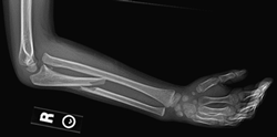
Fig. 1. Pediatric Monteggia fracture showing a proximal ulnar shaft fracture and a proximal radial dislocation.
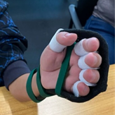
A study by Foran, et al., 2017 demonstrated that 83% of Monteggia fracture patients were successfully treated with conservative methods and did not require surgical intervention without compromising outcomes or increasing risk of complication. Typically, patients are immobilized either in a cast or custom thermoplastic long-arm orthotic for 4-6 weeks (Fig. 2).
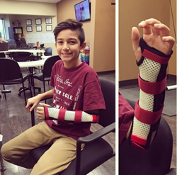
Fig. 2. Custom thermoplastic long-arm/ Muenster orthotic to support forearm and block full elbow range of motion.
Patients are highly monitored throughout the first three weeks, as this is the time period when instability is most likely to occur. If there is adequate healing of the ulna between 4-6 weeks, the cast is removed and the patient transitioned to removable forearm orthotic, at which time, therapy is initiated1.
Special considerations to monitor for:
- Compartment syndrome
- Gradual decreasing range of motion:
- Tendon/nerve injuries
- Skin breakdown
- Risk of recurrent fractures up to 6-12 months
Therapeutic interventions:
- Mobility to wrist/forearm
- Range of motion to all joints involved in orthotic
- Ideas include: painting on vertical surface, playing cards, tossing magnetic darts, wrist maze, sport simulation (overhead tossing, dribbling, racket movements, etc.)
- Building endurance
- Grip/pinch/lift strengthening and weight bearing
- Ideas include: animal walks for weightbearing, wall push-ups against yoga ball,
- Desensitization
- Over fracture site or in fingertips after nerve injury
- Ideas include: sensation kit (small squares of various materials transitioning from smooth to rough: velvet/moleskin, foam, Velcro, netting, sandpaper, etc.)
- Neuromuscular ed-education
- Re-training of movement patterns for ADL’s that are compromised from fracture involvement
- Ideas include: tendon glides, nerve glides, NMES for muscle activation
- Orthotics to prevent joint contractures & promote functional positioning
- Orthotics to protect over fracture site and prevent re-fracture
- Dynamic orthotics to support muscles groups weakened by neuropraxia/ injury
1. Foran, I., Upasami, V.V., Wallace, C.D., Britt, E., Bastrom, T.P., Bomar, J.D., & Pennock, A.T. (2017). Acute pediatric monteggia fractures: A conservative approach to stabilization. Journal of Pediatric Orthopedics, 37(6), 335-341.
More To Read

Hand therapy intervention activities for Chemo-Induced Peripheral Neuropathy (CIPN)
Blog Post Written By: Rita Steffes Patients with CIPN may present with symptoms that include numbness, tingling, hypersensitivity to cold, loss of tactile or vibration sensitivity, decreased balance, and shooting burning pain in their hands These symptoms make it difficult for oncology patients to participate in all activities of daily living with dressing, meal preparation,…

Intrinsic Hand Strengthening with Puttycise Tools
We are always looking for ways of the intrinsic hand strengthening. It is easy to overlook the importance of these small but mighty muscles. They are essential to performing functional grasps patterns. They can become weak in a short period of time due to their small size. So, How does intrinsic strengthening work?! The Basics…

Why Burnout Happens in Hand Therapy and What We Can Do About It.
Why Burnout Happens in Hand Therapy There are several reasons why burnout can occur, this is especially true for healthcare workers. What We Can Do About It Final Thought:Burnout isn’t a personal failure, it is often a systemic issue. But we do have power over how we respond. As hand therapists, we are experts at…

Mechanism of Interneural Edema in Carpal and Cubital Tunnel
Mechanism of Interneural Edema Over the last few weeks I have been learning about ultrasonic imaging and carpal tunnel syndrome. When reviewing carpal tunnel syndrome, I learned that intraneural edema is a common sign of compression injuries such as carpal tunnel and cubital tunnel. There are numerous causes of carpal tunnel syndrome, and every scenario…

Sign-up to Get Updates Straight to Your Inbox!
Sign up with us and we will send you regular blog posts on everything hand therapy, notices every time we upload new videos and tutorials, along with handout, protocols, and other useful information.