
Distal radius fracture types seen in the hand therapy clinic
Filed under Evaluation
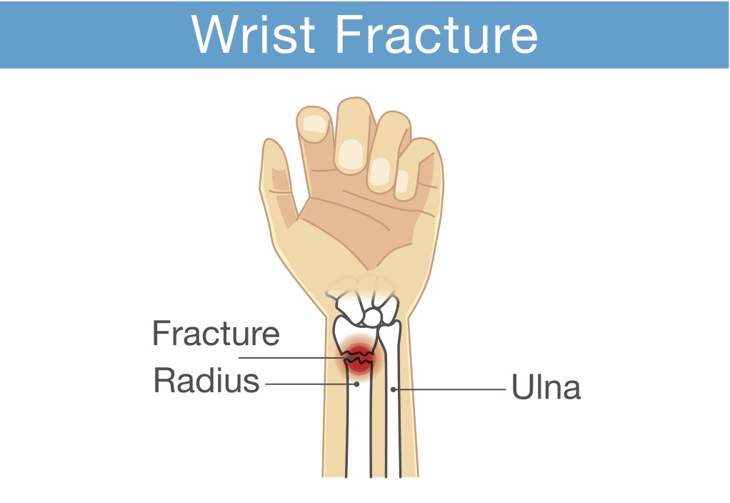
Distal radius fractures are one of the most common injuries seen in hand therapy. Several different distal radius fracture classification systems have been developed, and this blog post will focus on the more common types of distal radius fractures and their classification.
Extra-articular fractures are either nondisplaced or displaced fractures. These fractures occur outside of the joint.
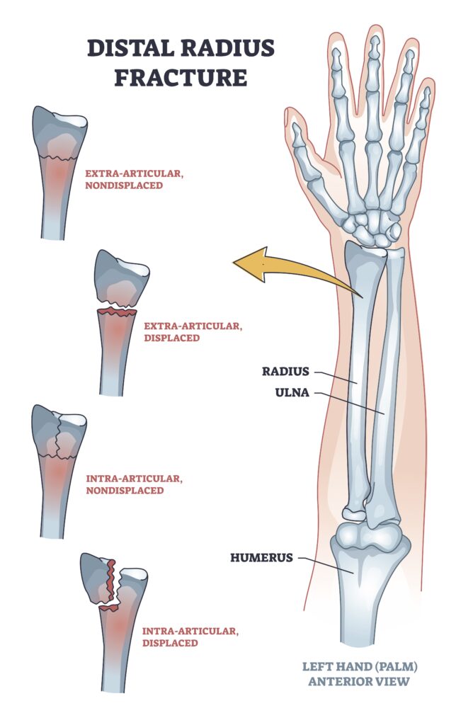
Similarly, Intra-articular fractures can be displaced or nondisplaced but occur within the joint.
Many fractures are named based on their fracture location, fracture pattern, and type of displacement.
Colles fractures are the most common type of distal radius fracture and account for about 90% of distal radius fractures. A Colles fracture is an extra-articular fracture with a dorsal displacement. These fractures occur from a fall forward on an outstretched hand.
A reverse Colles fracture is also known as Smith’s fracture. This is also an extra-articular fracture that is volarly displaced. These types of fractures are caused by falling backward and an outstretched arm.
Another type of distal radius fracture is a Barton’s fracture. This is an intra-articular fracture and is associated with a dislocation of the radio-carpal joint. A Barton’s fracture can be described as volar (more common) or dorsal (less common).
A die punch fracture is a depression fracture of the lunate fossa of the distal radius fracture that occurs with a vertical load through the lunate. These are often overlooked and not part of the classification system.
A Chauffeur’s fracture is also known as a radial styloid fracture or a Hutchinson’s fracture. This fracture is classified as an articular fracture. It was initially called a Chauffeur’s fracture because when the chauffeur would turn the crank to start the car, the motor often would cause the crank handle to jerk back.
1 Comment
Leave a Comment
More To Read

Prevention and Management of Upper Extremity injuries in Modern Mass Production
Injuries and Upper Extremty Pitts, G., Custer, M., Foister, R. D., & Uhl, T. (2021). The hand therapist’s role in the preventionand management of upper extremity injuries in the modern mass production industrial setting.Journal of Hand Therapy, 34(2), 237–249. https://doi.org/10.1016/j.jht.2021.04.019 By: Kaylen Kallander The Skinny: This study included four case studies to determine the impact…

Rapid Review: Is Finger Splinting Necessary after Flexor Tendon Repair?
Outcome of Flexor Tendon Repair Using Eight-Strand Core Stitch Without Postoperative Finger Splinting Reference: El-Gammal, T. A., Kotb, M. M., Ragheb, Y. F., El-Gammal, Y. T., & Anwar, M. M. (2024). Outcome of Flexor Tendon Repair Using Eight-Strand Core Stitch Without Postoperative Finger Splinting. HAND. https://doi.org/10.1177/15589447231220686 The Skinny: The purpose of this study was to…

Creating an Action Plan for Addressing Mental Health in the Clinic
Blog By: Rachel Reed As hand therapists, our care for our patients must be driven by the goal of treating the whole person, not just their hand or injury (Hannah, 2011). Occupational therapy is a unique profession in which we are equipped to view our patients through a holistic lens. With this lens, we are…

Pros and Cons of Cortisone Injections
By: Shruti Jani Patients will often times ask the therapist their opinion on cortisone injections. Cortisone injections can be very helpful and significantly reduce inflammation, however, some therapists feel this can mask the pain not treating the true root cause of the problem. This is often debated among therapists. A short synopsis of the pros…

Sign-up to Get Updates Straight to Your Inbox!
Sign up with us and we will send you regular blog posts on everything hand therapy, notices every time we upload new videos and tutorials, along with handout, protocols, and other useful information.
Thank you so much! I always enjoy your informative blogs and have learned a great deal. Your effort is very much appreciated!!!