
Distal radius fracture types seen in the hand therapy clinic
Filed under Evaluation
Distal radius fractures are one of the most common injuries seen in hand therapy. Several different distal radius fracture classification systems have been developed, and this blog post will focus on the more common types of distal radius fractures and their classification.
Extra-articular fractures are either nondisplaced or displaced fractures. These fractures occur outside of the joint.
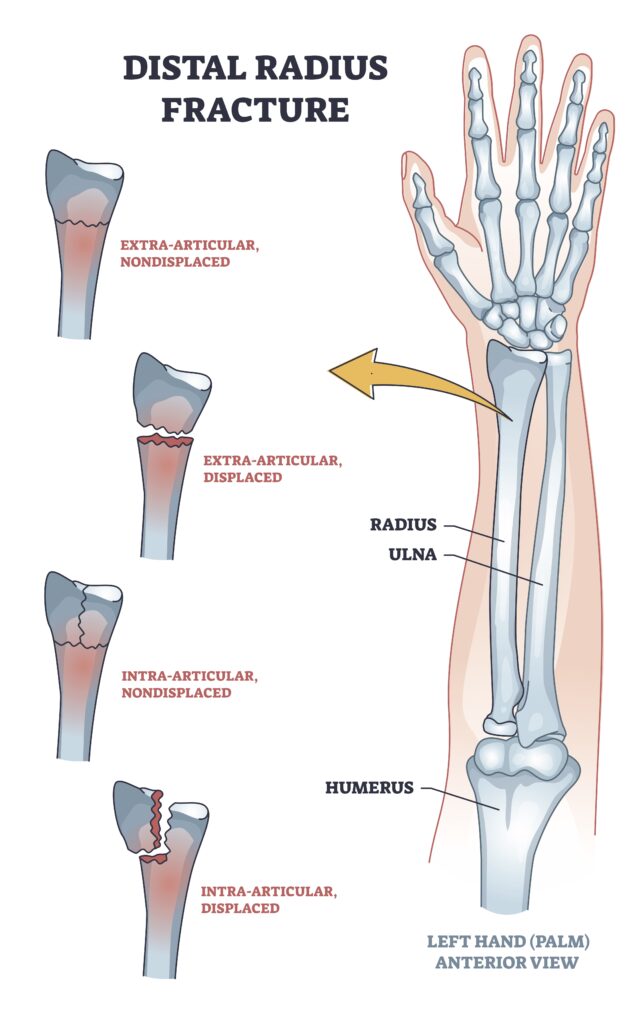
Similarly, Intra-articular fractures can be displaced or nondisplaced but occur within the joint.
Many fractures are named based on their fracture location, fracture pattern, and type of displacement.
Colles fractures are the most common type of distal radius fracture and account for about 90% of distal radius fractures. A Colles fracture is an extra-articular fracture with a dorsal displacement. These fractures occur from a fall forward on an outstretched hand.
A reverse Colles fracture is also known as Smith’s fracture. This is also an extra-articular fracture that is volarly displaced. These types of fractures are caused by falling backward and an outstretched arm.
Another type of distal radius fracture is a Barton’s fracture. This is an intra-articular fracture and is associated with a dislocation of the radio-carpal joint. A Barton’s fracture can be described as volar (more common) or dorsal (less common).
A die punch fracture is a depression fracture of the lunate fossa of the distal radius fracture that occurs with a vertical load through the lunate. These are often overlooked and not part of the classification system.
A Chauffeur’s fracture is also known as a radial styloid fracture or a Hutchinson’s fracture. This fracture is classified as an articular fracture. It was initially called a Chauffeur’s fracture because when the chauffeur would turn the crank to start the car, the motor often would cause the crank handle to jerk back.
1 Comments
Leave a Comment
More To Read

Tennis Elbow and Graded Exercises
Lateral Elbow Pain with Graded Exercise Chronic tennis elbow with a supervised graded exercise protocol Özdinçler, A. R., Baktır, Z. S., Mutlu, E. K., & Koçyiğit, A. (2023). Chronic lateral elbow tendinopathy with a supervised graded exercise protocol. Journal of Hand Therapy, 36(4), 913–922. https://doi.org/10.1016/j.jht.2022.11.005 The Skinny: This study looked at the effectiveness of an…
Read MoreDoes Taking an Alpha-lipoic for 40 days after Carpal Tunnel Release decrease the likelihood of developing Pillar Pain?
Filippo, B., Granchi, D., Roatti, G., Merlini, L., Sabattini, T., & Baldini, N. (2017). Alpha-lipoic acid after median nerve decompression at the carpal tunnel: A randomized controlled trial. The Journal of Hand Surgery, 4, 236–42. The Skinny – A double-blind, randomized controlled study was performed. Sixty-four patients were randomly assigned into two groups after median…
Read More
Dorsal Scapular Nerve Entrapment and Thoracic Pain
Don’t Forget to Evaluate for Dorsal Scapular Nerve Entrapment By Delaney Wright If your patient presents with any upper thoracic pain, it is critical to take measures to evaluate for dorsal scapular nerve entrapment. In a study completed by Sultan et al. (2013), 55 patients with interscapular pain were evaluated clinically and via nerve conduction…
Read More
Sign-up to Get Updates Straight to Your Inbox!
Sign up with us and we will send you regular blog posts on everything hand therapy, notices every time we upload new videos and tutorials, along with handout, protocols, and other useful information.
Thank you so much! I always enjoy your informative blogs and have learned a great deal. Your effort is very much appreciated!!!