

Crowley, J. S., Meunier, M., Lieber, R. L., & Abrams, R. A. (2020). The Lumbricals Are Not the Workhorse of Digital Extension and Do Not Relax Their Own Antagonist. The Journal of Hand Surgery.
The Skinny:
What do the lumbricals do?
There is a long-standing belief that the lumbricals act as a counterforce to the digital flexors enough to relax the digital flexors and act as primary digital extensors. This article works to update our understanding of the role of the lumbrical based on anatomy and innervation. They build off the work of Wang et al. to develop a better understanding of what the lumbricals function actually is in digit extension and balance of forces.
In The Weeds:
To review the anatomical line of the lumbricals, check out the picture below. Also, the lumbricals have several unique properties. They have the highest spindle fiber density of any UE muscle (a property of muscles intended for refined graded control, not power). They also have the highest fiber length-to-muscle length ratio. This means that the lumbricals are best at providing a low amount of force over a wide range of motion. These two aspects refute the idea that the lumbricals are strong enough to overpower counterbalance the much larger, conversely designed, FDP & FDS.
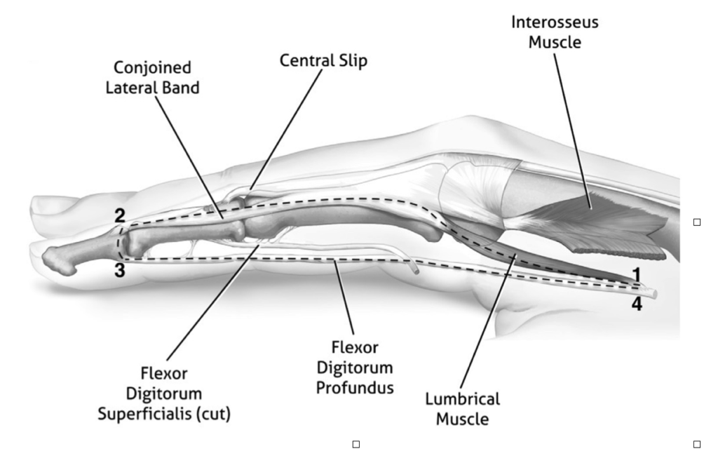
Crowley, J. S., Meunier, M., Lieber, R. L., & Abrams, R. A. (2020). The Lumbricals Are Not the Workhorse of Digital Extension and Do Not Relax Their Own Antagonist. The Journal of Hand Surgery.
Bringing It Home:
So what? What does that mean? Rather than the lumbricals being some powerhouse of IP extension, this article builds on the Wang articles proposal. Want et al. propose that the lumbricals are a tension monitoring device that allows for refined motor coordination to aid in coordinating the stronger extrinsic muscles. Crowley et al. add to that and suggest that the lumbricals also serve as a spring in a closed loop that helps to balance the 2nd and 3rd phalanx over the 1st, since the 1st phalanx has no tendon insertions to maintain its position. That makes the 1st phalanx an intercalated segment that needs delicately maintained tension to preserve its position. This is a significant shift in the idea that the lumbricals are a strong force in digital extension, counteracting the FDP & FDS.
Rating: 5/5
While not a research study, this article adds a lot of understanding and clarification of the anatomy and role of the lumbricals. Looking at muscle size, length, fiber type, and anatomical attachment can improve our awareness of a unique muscle like this is actually doing in the hand. It is always good to stay open-minded to alternative views of our sometimes dogmatically held opinions and beliefs. There are many more significant pieces of info in this article, and I highly recommend reading it if you have a chance.
Wang K, McGlinn EP, Chung KC. A biomechanical and evolutionary perspective on the function of the lumbrical muscle. J Hand Surg Am. 2014;39(1):149e155.
1 Comment
Leave a Comment
More To Read
Which orthosis design is better for zone 5-6 extensor tendon injuries, a relative motion orthosis compared to a dynamic extension orthosis?
M. Buhler, ˝ D. Gwynne-Jones, M. Chin et al., (2023) Are the outcomes of relative motion extension orthoses noninferior and cost-effective compared with dynamic extension orthoses for management of zones V-VI finger extensor tendon repairs: A randomizedcontrolledtrialJournalofHandTherapy. The Skinny: The aim of this study was to compare the data for two different types of orthoses…

Do you know the secret ingredient to recovering from an injury?
Do you know the secret ingredient to recovering from an injury? I will give you a hint it is 5 letters and begins with the letter S. SLEEP Have you ever asked yourself a question – does sleep help injuries heal? This is for you to share with your patients but also serve as a…

Biceps Tenodesis Versus Tenotomy During Arthroscopic Rotator Cuff Repair
Article Review By: Delaney Wright Title: Outcomes of Biceps Tenodesis Versus Tenotomy During Arthroscopic Rotator Cuff Repair: An Analysis of Patients From a Large Multicenter Database Reference: Srinivasan, R. C., Hao, K. A., Wright, T. W., Farmer, K. W., Wright, J. O., Roach, R. P., Moser, M. W., Freidl, M. C., Pazik, M., & King,…

Wrist Proprioception Ideas for Hand Therapy
Wrist Proprioception Intervention Ideas: By Ammie Ingwaldson Lack of wrist proprioception exercises can affect clients in the hand therapy setting with neurological and musculoskeletal conditions. Proprioception limitations are found in common conditions such as carpal tunnel syndrome, distal radius fracture, and CRPS (Valdes, Naughton & Algar, 2014). Proprioception is necessary during daily tasks to provide…

Sign-up to Get Updates Straight to Your Inbox!
Sign up with us and we will send you regular blog posts on everything hand therapy, notices every time we upload new videos and tutorials, along with handout, protocols, and other useful information.
Great explanation and illustration