
Mechanism of Interneural Edema in Carpal and Cubital Tunnel
Filed under Diagnoses
Mechanism of Interneural Edema
Over the last few weeks I have been learning about ultrasonic imaging and carpal tunnel syndrome. When reviewing carpal tunnel syndrome, I learned that intraneural edema is a common sign of compression injuries such as carpal tunnel and cubital tunnel. There are numerous causes of carpal tunnel syndrome, and every scenario ends with the reduction of available space within the carpal tunnel and the inevitable compression on the median nerve (carpal tunnel edema). What I did not know was that chronic compression on the nerve can disrupt and open the blood nerve barrier around the perineurial layer. This allows for blood to flow freely into the nerve causing swelling or interneural edema. Since the nervous system lacks lymphatic drainage in the endoneural space, swelling inevitably increases pressure and disrupts the flow of blood to the nerve resulting in a metabolic conduction block (Cooper, 2014). One animal study found that an increase in pressure as little as 30 grams of force (about the weight of an average lightbulb) over the course of 1 hour was enough to disrupt the blood nerve barrier around the median nerve and cause diffusion (Kobayashi et al., 2005).
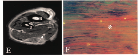
Normal Nerve No Diffusion
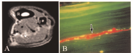
Nerve 30 Grams of Force with Diffusion
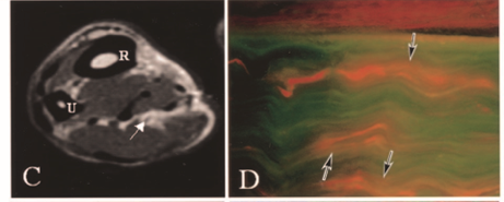
Nerve 90 Grams of Force Severe Diffusion
Chronic compression and decreased blood flow lead to impairment in nerve conduction. One source states that functional deficits are seen sequentially in the following order: motor, proprioception, touch, temperature, pain, and then sympathetic function (Cooper, 2014). Therapeutic activities such as nerve gliding exercises are hypothesized to increase nerve mobility and release the nerve from the sight of compression. Additionally, surgical decompression can help to alleviate symptoms, but the timeline for neural repair is largely based on the severity of nerve damage that has occurred. As neural edema subsides and blood flow to the nerve improves, the nerve begins to repair itself as long as the endoneurial tubes are intact. Patients are expected to regain sensation in the reverse order that they were initially lost (pain, temperature, proprioception).
Cooper, C. (20014). Fundamentals of hand therapy: Clinical reasoning and treatment guidelines for common diagnoses of the upper extremity [Second Edition]. Elsevier Mosby
Kobayashi, S., Meir, A., Baba, H., Uchida, K., and Hayakawa, K. (2005). Imaging of intraneural edema by using gadolinium-enhanced MR imaging: Experimental compression injury
2 Comments
Leave a Comment
More To Read

Management of the Stiff Finger: Evidence and Outcomes
Title: Management of the Stiff Finger: Evidence and Outcomes Reference: Yang, G., McGlinn, E. P., & Chung, K. C. (2014). Management of the stiff finger: Evidence and outcomes. Clinics in Plastic Surgery, 41(3), 501–512. https://doi.org/10.1016/j.cps.2014.03.011 Article Review By: Tommi Hintnaus The Skinny: This study focuses on understanding and managing finger stiffness, a condition characterized by…
Read More
A Fun Fact from a Hand Therapy Student
By: Ammie Ingwaldson Level 2 Fieldwork at a hand therapy clinic is a fast paced and continuous learning experience. The perfect example of this occurred last week while observing a therapist provide a client with their home CMC arthritis program. The therapist was educating the client on how to oppose their thumb to their small…
Read More
Hand Therapy Marketing 101
Marketing 101 – 5 Tips for Your Therapy Clinic Confession: I hate marketing. It’s my least favorite part of my job. It is so hard to open yourself up to that much rejection but still stay positive. It feels like the professional version of blind dating, except the other person probably already has a significant…
Read More
Sign-up to Get Updates Straight to Your Inbox!
Sign up with us and we will send you regular blog posts on everything hand therapy, notices every time we upload new videos and tutorials, along with handout, protocols, and other useful information.
The inter neural edema is fascinating.
Yes is is very interesting! I agree