
Video-augmented mirror therapy for upper extremity rehabilitation after stroke
Filed under Treatments
Kim, H., Kim, J., Jo, S., Lee, K., Kim, J., & Song, C. (2023). Video augmented mirror therapy for upper extremity rehabilitation after stroke: a randomized controlled trial. Journal of Neurology, 270(2), 831-842.
Article Review: Shannon Skowbo
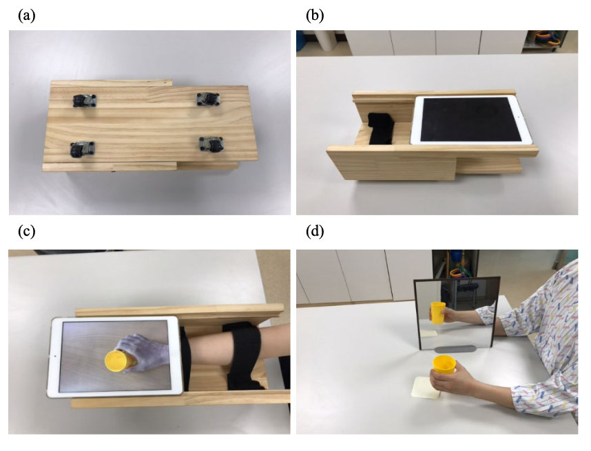
The Skinny: This single-blind, randomized control trial aimed to assess the effects of mirror therapy for stroke patients using a video-augmented device (MTVAD) on reach-to-grasp tasks and upper extremity motor function compared to traditional mirror therapy (TMT). TMT has some limitations. It requires bilateral movements since the unaffected limb produces the illusion, promotes asymmetrical postures to allow for viewing the reflection, and has discrepancies between movement performance and visual feedback. Kim et al. (2023) explores a novel therapeutic method using MTVAD to target the aforementioned limitations of TMT.
In the Weeds: Inclusion criteria included first-time stroke patients with hemiplegia during the previous 12 months, mild to moderate motor impairment as determined by upper extremity scores of 26-56 on the Fugel Myer Assessment (FMA), ability to understand and follow simple directions, and a score of greater than or equal to 21 on the Korean version of the Mini-mental State Examination. Exclusion criteria included psychiatric disorders or dementia, orthopedic disorders, apraxia or hemineglect, and prior experience with mirror therapy. Participants were randomly assigned to one of three groups: MTVAD, TMT, or the control group (conventional rehabilitation). MTVAD and TMT were performed for 30 minutes per day, 5 times per week, for 4 weeks. The control group received conventional rehabilitation for 60 minutes per day, 5 times per week, for 4 weeks. MTVAD and TMT received the same amount of conventional rehabilitation as the control group. The outcome measures included a variety of kinematic parameters during a reach-to-grasp task, upper extremity subscores on the FMA, and upper extremity subscores on the Manual Function Test (MFT). 36 participants were included in the statistical analysis.
Bringing it Home: The MVTAD group showed significantly greater improvements than the TMT and control groups in movement time, peak velocity, and trunk displacement. This means that the MVTAD group could perform the reach-to-grasp tasks more quickly, efficiently, and with a straighter trunk. The MVTAD also showed significantly greater improvements than the TMT group in FMA subscores for the shoulder, elbow, and forearm and MFT subscores for the shoulder. The improvements in kinematic parameters suggest that MTVAD “promoted better performance and upper extremity motor control ability during the reach-to-grasp movement compared to TMT in patients with stroke” (Kim et al., 2023, p. 838).
Rating: Overall, this study received a 4/5 rating. The pre-and post- test blinding, randomization, and clinically significant sample population size indicate the study has strong internal validity. However, these results can only be generalized to stroke patients who are within one year from their first stroke with only mild to moderate motor deficits.
More To Read

What is the incidence of musculoskeletal complaints in the elbow, shoulder, and neck after hand and forearm injuries?
Winiarski, L. M., Livoni, J. D., Madsen, P. V., Rathleff, M. S., & Larsen, P. (2021). Concurrent musculoskeletal complaints in elbows, shoulders, and necks after common hand and forearm injuries or conditions: A cross-sectional study among 600 patients. Journal of hand therapy: official journal of the American Society of Hand Therapists, 34(4), 543–548. https://doi.org/10.1016/j.jht.2020.05.002 The Skinny: The…

Simple but Effective Ways Hand Therapists Address Psychosocial Impacts of Upper Extremity Injuries
Although psychosocial factors are often not formally assessed during an evaluation in those with upper extremity injuries, the therapist often informally assesses these during and after treatment sessions. Sustaining an upper extremity injury can be a physically and emotionally challenging experience. Beyond the physical pain and limitations, these injuries can profoundly impact an individual’s psychosocial…

Tennis Elbow and Graded Exercises
Lateral Elbow Pain with Graded Exercise Chronic tennis elbow with a supervised graded exercise protocol Özdinçler, A. R., Baktır, Z. S., Mutlu, E. K., & Koçyiğit, A. (2023). Chronic lateral elbow tendinopathy with a supervised graded exercise protocol. Journal of Hand Therapy, 36(4), 913–922. https://doi.org/10.1016/j.jht.2022.11.005 The Skinny: This study looked at the effectiveness of an…

Shoulder Pain: The Effectiveness of Conservative Treatment
Reference: Steuri, R., Sattelmayer, M., Elsig, S., Kolly, C., Tal, A., Taeymans, J., & Hilfiker, R. (2017). Effectiveness of conservative interventions including exercise, manual therapy and medical management in adults with shoulder impingement: a systematic review and meta-analysis of RCTs. British journal of sports medicine, 51(18), 1340–1347. https://doi.org/10.1136/bjsports-2016-096515 By: Tayler Roost The Skinny: This study…

Sign-up to Get Updates Straight to Your Inbox!
Sign up with us and we will send you regular blog posts on everything hand therapy, notices every time we upload new videos and tutorials, along with handout, protocols, and other useful information.