
Pillar Pain After Carpal Tunnel Release Surgery
Carpal tunnel release (CTR) surgery is a common procedure, with the majority of patients experiencing satisfaction with its outcomes. However, for some individuals, a temporary complication known as “pillar pain” may arise, affecting approximately 13% of those undergoing CTR.

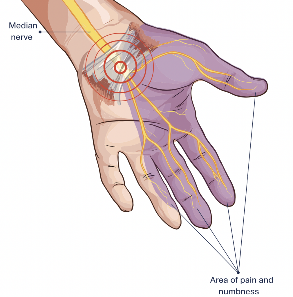
Pillar pain manifests in the thenar eminence and hypothenar eminence due to their proximity to the transverse carpal ligament. It is characterized by wrist pain that causes tenderness and discomfort upon touch, distinct from the incision pain typical of surgical recovery, which usually subsides within a few days to weeks.
Although pillar pain symptoms typically resolve within three months, they can persist longer, even up to 9-12 months. While the exact cause of pillar pain remains elusive, several theories have been proposed, including tender scar tissue, muscle alignment alterations, joint inflammation, injury to small nerve fiber branches, and nerve irritation. Notably, a neurogenic component is suggested, indicating that nerve tissue damage from carpal tunnel surgery may play a significant role.
A theory posits that avoiding the “critical pillar rectangle” during surgery, which encompasses specific anatomical landmarks, could substantially reduce the likelihood of pillar pain occurrence.
Hand therapists offer various interventions for managing pillar pain, including:
- Stretching exercises such as the extrinsic stretching of wrist/hand flexor and tendon gliding exercises.
- Soft tissue mobilization targeted at the thenar and hypothenar eminences, gradually increasing pressure in sensitive areas.
- Desensitization techniques involve the use of different textures like cotton, wool, foam, soft Velcro, and others to rub over sensitive areas. Immersion of the affected area in substances like cotton balls, foam, rice, or beans can aid in desensitization.
- Median nerve glides to facilitate nerve flossing and restore median nerve mobility.
- Scar softening treatments such as paper tape or silicone gel pad.
By employing these strategies, individuals experiencing pillar pain can effectively manage their symptoms and facilitate recovery following carpal tunnel surgery.
Kumar, A., & Lawson-Smith, M. (2024). Pillar pain after minimally invasive and standard open carpal tunnel release: A systematic review and meta-analysis. Journal of Hand Surgery Global Online. https://doi.org/10.1016/j.jhsg.2023.12.003
Ludlow, K., Merla, L., Cox, J., & Hurst, L. (1997a). Pillar pain as a postoperative complication of carpal tunnel release. Journal of Hand Therapy, 10(4), 277–282. https://doi.org/10.1016/s0894-1130(97)80042-7 .
2 Comments
Leave a Comment
More To Read
")
When should you use a Static Progressive Splint in Hand Therapy?
Flowers, K. (2002). A proposed decision hierarchy for splinting the stiff joint, with an emphasis on force application parameters. Journal of Hand Therapy, 15, 158–162. The Skinny- The article proposes a decision hierarchy to determine when you should apply a static progressive or dynamic orthosis. The decision hierarchy uses a modified Weeks test (MWT). The…

The Use of Neuromuscular Electrical Stimulation with Upper Extremity Paralysis
The Use of Neuromuscular Electrical Stimulation with Upper Extremity Paralysis By: Mikayla Murphy Martin, R., Johnston, K., & Sadowsky, C. (2012). Neuromuscular electrical stimulation–assisted grasp training and restoration of function in the tetraplegic hand: A case series. The American Journal of Occupational Therapy, 66(4), 471-477. https://doi.org/10.5014/ajot.2012.003004 The Skinny The purpose of the study was to…

Orthotic Options for Hand Burns
By: Sophia Grimm Hand burns can be very challenging to treat, and successful rehabilitation begins early after acute injury. Following a burn injury, scar contractures are the primary reason for the deformity of the hand. Therefore, proper orthotic intervention is key to preventing joint and ligament contractures (Kelly, Berenz & Williams, 2019). Splinting goals following…

Handlebar Palsy also known as Ulnar Nerve Compression
Handlebar Palsy also known as Ulnar Nerve Compression Handlebar palsy, also known as ulnar nerve compression, is a condition commonly experienced by cyclists due to prolonged pressure on the ulnar nerve at the wrist in an area called Guyon’s Canal. This pressure can occur from putting pressure on the handlebars or gripping the handlebars tightly. …

Sign-up to Get Updates Straight to Your Inbox!
Sign up with us and we will send you regular blog posts on everything hand therapy, notices every time we upload new videos and tutorials, along with handout, protocols, and other useful information.
Thank you for all the info
Great information, thanks! 🙏🏻