
By Brittany Carrie
A Student’s Perspective
During the first few weeks of my rotation, I was exposed to many new and exciting things that I had not been exposed to in the classroom setting. I observed and helped treat patients who had undergone severe trauma from lacerating tendons to complete amputations, saw different splinting techniques, and learned about some rather unique cases that are often characterized by significant pain with no physical representation on X-ray or other diagnostic imaging. One day, one of our patients who has had significant pain and discomfort in her fingers brought in a remarkable X-ray that, to my eyes, appeared flawless! I could see every bone so vividly; it was unlike any other X-ray image I had seen in a textbook! However, located near the MCP of the patient’s thumb in both hands was a small round bone that I can only describe as looking like a small seed in the hand. I was unsure what this bone was, and my mentor told me it is a sesamoid bone (sesamoid bone thumb)! This led me to further investigation. I wanted to know what sesamoid bones were, what they do and how a sesamoid bone hand therapy is done, BUT through my research, it appears researchers still do not know exactly what they do or why they exist. The following provides some general themes I found through a small literature search.
To begin with, sesamoid bones in hand are small, oval shaped bones that resemble the appearance of sesame seeds (Koo, Song, Sung, Lee, & Jun, 2017; Yammine, 2014; Yammine 2018). They are found within tendons primarily in the hands and feet and are seen radiographically. There are sesamoid bones found in other locations as well including the patella and fabella in the knee and the pisiform on the wrist (Yammine, 2014).
In most cases, sesamoid bones are found within the MCP joint of the thumb, index, and small fingers; however, the number of sesamoids at each joint varies across gender and population (Koo, Song, Sung, Lee, & Jun, 2017; Yammine, 2014; Yammine 2018). Figure 1 illustrates sesamoid bones located in the MCP and IP joints. In a study done by Yammine (2014), it was found that women often have more sesamoid bones in their hands compared to men. Most people typically have 5 sesamoid bones in each hand—the thumb has 2 in the MCP joint and 1 in the IP joint; the index finger has 1 in the MCP joint; the small finger has 1 in the MCP (Yammine, 2014; Yammine, 2018). However, research has shown that the East Asian and European populations have a higher amount of sesamoid bones in the hand compared to African and Middle Eastern populations, and the Japanese have a higher amount of sesamoid bones in comparison to the Chinese (Koo, Song, Sung, Lee, & Jun, 2017; Yammine, 2014).
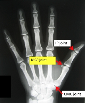
Fig 1. Radiographic image of sesamoid bones located at MCP and IP joints. Retrieved from http://www.myhand.com.au/m/index.php?option=com_content&view=article&id=50&Itemid=128&font-size=larger
The presence and function of sesamoid bones still remains unclear; however, there are a few hypotheses that include both functional and phylogenetic ideas. The functional hypothesis presents the idea that sesamoid bones increase tendon leverage to decrease friction and alter the direction of muscle action which increases pinch strength and MCP range of motion (Koo, Song, Sung, Lee, & Jun, 2017). Sesamoids are also believed to assist with joint stability and provide capsular strengthening (Yammine, 2014). The phylogenetic hypothesis describes the presence of sesamoid bones beginning during embryo development (Koo, Song, Sung, Lee, & Jun, 2017; Yammine, 2014). In sum, sesamoid bones are a unique feature to the human body that still remain a mystery as to what they do, why they exist, and why they are more prominent in certain populations.
Koo, B. S., Song, Y., Sung, Y.-K., Lee, S., & Jun, J.-B. (2017). Prevalence and distribution of sesamoid bones in the hand determined using digital tomosynthesis. Clinical Anatomy (New York, N.Y.), 30(5), 608–613.
Yammine, K. (2014). The prevalence of the sesamoid bones of the hand: A systematic review and meta-analysis. Clinical Anatomy (New York, N.Y.), 27(8), 1291–1303.
Yammine, K. (2018). The relationship between digit independence and digital sesamoids in humans and a proposal of a new digital sesamoid evolutionary hypothesis. Anatomical Record (Hoboken, N.J.: 2007), 301(6), 1046–1060.
15 Comments
Leave a Comment
More To Read

Is HEP Just as Good as Therapy for Metacarpal Fracture Rehab?
Gülke, J., Leopold, B., Grözinger, D., Drews, B., Paschke, S., & Wachter, N. J. (2018). Postoperative treatment of metacarpal fractures – Classical physical therapy compared with a home exercise program. Journal of Hand Therapy, 31(1), 20-28. The Skinny – Medicine is moving towards a model that encourages less direct intervention and a more DIY focus…

How To Do A Fast but Thorough Hand Therapy Assessment
We don’t get a lot of time. Sometimes new patients come in unexpectedly or someone comes at the wrong time and your 1-hour block for an eval is suddenly only 30 minutes. Do you know how to get the most out of your eval time with the patient? Do you know what things are the…

3 Common Reasons for Ulnar-Sided Wrist Pain and Non-Surgical Hand Therapy Treatment Options
3 Common Reasons for Ulnar Sided Wrist Pain

Differential Diagnosis: Trigger Finger vs. Subluxing Sagittal Band Injury vs. Subluxing Lateral Band
Differential Diagnosis: Trigger Finger vs. Subluxing Sagittal Band Injury vs. Subluxing Lateral Band Hand therapists frequently encounter patients presenting with finger pain, clicking, and difficulty with tendon glide. Among the most commonly confused conditions are trigger finger, subluxing sagittal band injury, and subluxing lateral band. Each of these pathologies involves different anatomical structures and biomechanical…

Sign-up to Get Updates Straight to Your Inbox!
Sign up with us and we will send you regular blog posts on everything hand therapy, notices every time we upload new videos and tutorials, along with handout, protocols, and other useful information.
Fascinating and helpful. Thank you so much for putting this together.
Thank you! It was an interesting read!
Very interesting. In a CHT and I learned something new!! I didn’t know there were so many in the different i joints other than thumb IPJ . I definitely didn’t know difference in hereditary differences?
Thanks for the note! I found it very interesting as well. It makes you wonder what all the clinical implications are.
Miranda
Since there’s really only 2 research authors (Yammine and Koo, et. al), I kind of worry about the validity of these findings – and if there’s possibly some cultural elitism… Just saying…
It sounds like you are assuming that more sesamoid bones is indicative of advancement/superiority. Is it possible that the increased presence of sesamoid bones is actually a sign of dysfunction? To me it would seem that nuggets of bone growing within tendons would indicate excessive loads on the tendon, probably as a result of compensating for structural misalignment elsewhere in the body, such as the spine or pelvis. Based on the cultural frequencies described in the post, my first reaction is that sesamoid frequency correlates with technological advancement. Cultures that have offloaded the use of the body to machines tend to have increased sedentism, which expedites the deterioration of the body, including the musculoskeletal system. The presence of an above-average number of sesamoid bones could merely be a symptom of this kind of systemic dysfunction.
I recently suffered a 100% MCP dislocation because of a car accident (airbag). The x-rays show a fractured sesamoid, and I wanted to learn more about this little bone. Thank you for gathering this information!
How did you treat it?
This was really interesting. I broke my arm and saw the sesamoid on the x-ray. The ortho doc explained what it was but couldn’t remember the nane for it and said she had never been told why we had them. So, I poured over undergrad and grad anatomy and physiology books and I couldn’t find anything about them. Thanks for doing such a good job providing references, too. I don’t think I will ever forget this cool anatomy fact now. Yea sesamoids!
had an x-ray done on both hands, and on both hands on the index, pointer, and thumb have these sesamoid bones! Doc said I’m symmetrical! I had no idea of these “extra” bones in our bodies, pretty cool, even cooler since we’re unaware of most of it today. Thanks for the read
They’re “cool” only if they behave and don’t cause pain (which mine does). Cortisone shots help, if done right, but don’t last very long. Only option is removal, which the doctor said to put off as long as possible because of loss of strength in the thumb. It’s a “rock and a hard place” situation.
Thank you for sharing.
I forgot to tell the author Thank You for your article on this. I’ve not found anything else to explain it as well as you did.
Thank you for your kind words!
I tore the MCP 2yrs ago, & the Sesamoid was not there. I never got it fixed for various reasons i could not contol. But i tripped & fell 2 days ago, & todays xray showed the bigger of the two Sesamoid bones out of place now. Unfortunately this time i WILL need to get the thumb repaired. it’s going to hurt.