
Glenohumeral Joint Ligaments
The Glenohumeral (GH) joint is composed of the head of the humerus and the glenoid fossa. The fossa is relatively small compared to the humeral head, making the joint highly mobile, which also leads to an increased risk of instability.
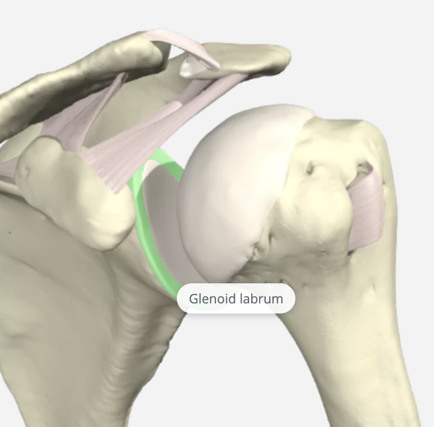
The glenoid labrum is a fibrocartilagenous rim attached around the glenoid that helps deepen the glenoid fossa by 50%, providing increased stability of the GH joint.
The GH joint relies heavily on the soft tissue structures for stability, and the GH ligaments are the primary static stabilizers of the joint. These include the Coracohumeral Ligament (CHL), Superior Glenohumeral Ligament (SGHL), Middle Glenohumeral Ligament (MGHL), Inferior Glenohumeral Ligament (IGHL), and the Posterior Inferior Glenohumeral Ligament (PIGHL).
Coracohumeral Ligament (CHL)
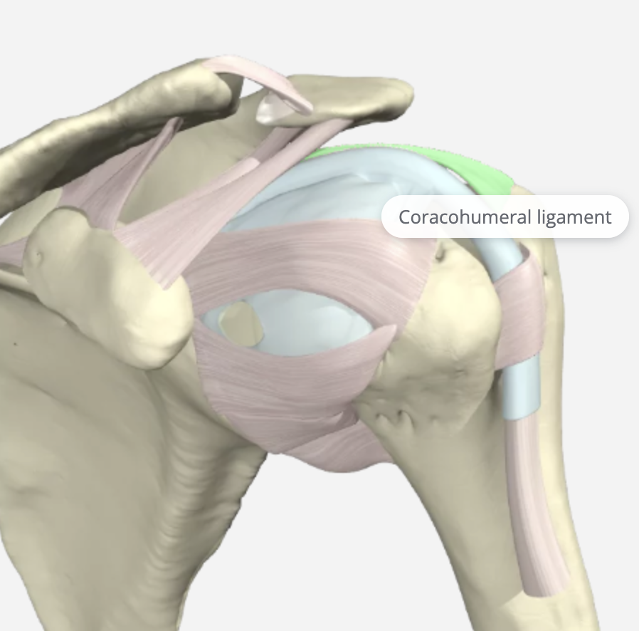
The CHL prevents superior dislocation and inferior displacement of the humerus. It is included in this review as it blends with the (SGHL). It is divided into two parts, the anterior and posterior bands. The Anterior Coracohumeral Ligament inserts on the lesser tuberosity and is tight in 30 degrees shoulder extension. The Posterior Coracohumeral Ligament inserts on the greater tuberosity and tight flexion at 60-70 degrees. It is also a secondary restraint in preventing the long head of the biceps from subluxing medially.
Superior Glenohumeral Ligament (SGL)
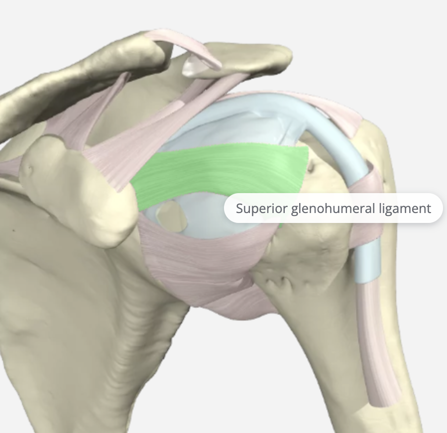
The SGHL is the smallest and least understood ligament in the GH capsule. Its origin is the upper part of the glenoid cavity and the base of the coracoid process. It attaches to the MGL, the biceps tendon, and the labrum. It is tight in adduction, the middle at 45 degrees of abduction, and when the shoulder is brought to 90 degrees of abduction with external rotation. It works with the CH ligament to prevent inferior translation of the humeral head.
Middle Glenohumeral Ligament (MGHL)
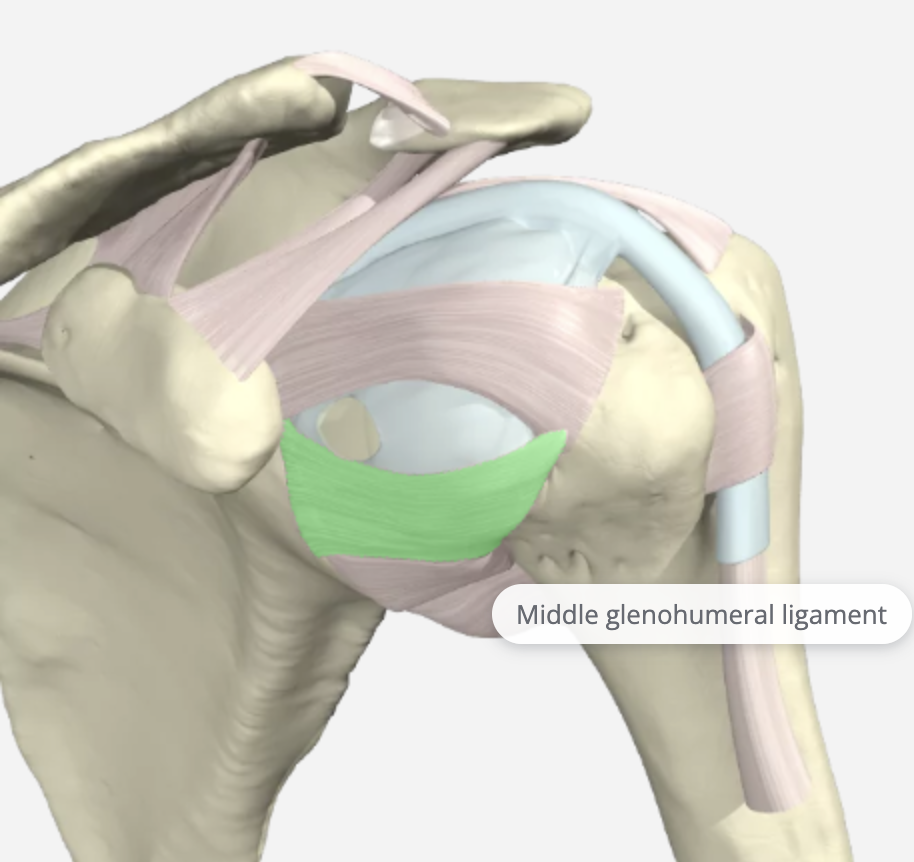
The middle glenohumeral ligament (MGHL) attaches to the anterior aspect of the anatomic neck of the humerus, just medial to the lesser tuberosity. It arises from the glenoid by way of the labrum. Of the three glenohumeral ligaments, the MGL demonstrates the most significant variation in size. It is tight in the abduction and provides anterior stability at 45 degrees and 60 degrees abduction. Injuries to this area alone are very rare and are never isolated.
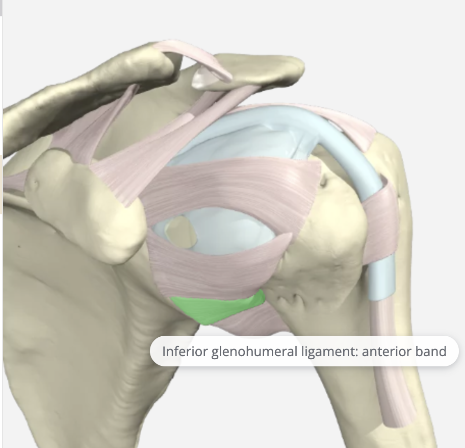
Inferior Glenohumeral Ligament (IGHL)
IGHL is tight in true abduction and slightly looser in the scapular plane of abduction. It originates from the glenoid labrum and inserts into the humeral neck. It is the most important stabilizer against anterior-inferior shoulder dislocation. Therefore this component is the most frequently injured and is most likely to tear when the arm is fully abducted. It is the strongest and most important soft tissue stabilizer. It can be avulsed from the glenoid side resulting in an anteroinferior labral tear.
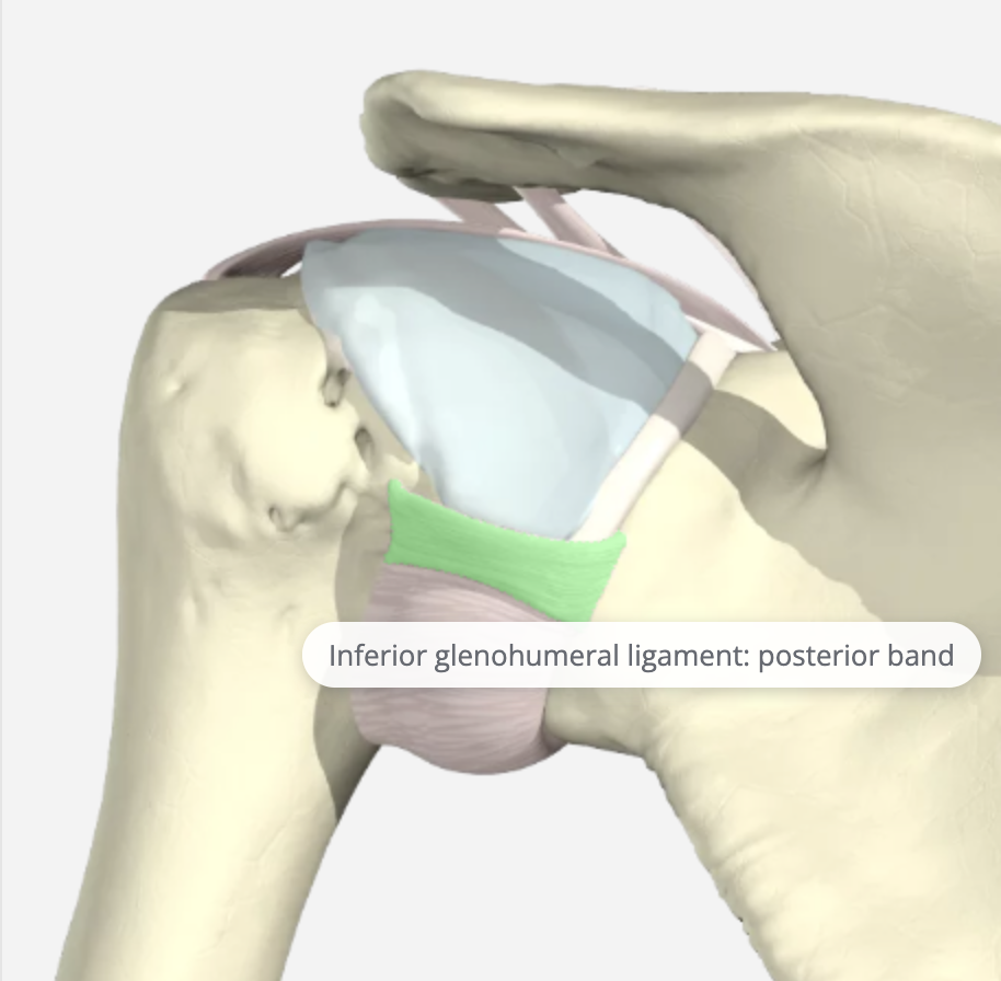
Posterior Inferior Glenohumeral Ligament (PIGHL)
PIGHL is not as robust as the anterior ligaments but is essential to balance the capsule. Laxity in this part of the capsule is considered normal. The posterior band of the IGLC is mainly responsible for capsuloligamentous restraint to posterior translation of humeral head in 90° of abduction.
Goetti, P., Denard, P. J., Collin, P., Ibrahim, M., Hoffmeyer, P., & Lädermann, A. (2020). Shoulder biomechanics in normal and selected pathological conditions. EFORT open reviews, 5(8), 508–518. https://doi.org/10.1302/2058-5241.5.200006
1 Comment
Leave a Comment
More To Read

How to use Kinesiology Taping for Shoulder Subluxation
How to us Kinesiology Tape for Shoulder Subluxation By: Tayler Roost What is shoulder subluxation? Shoulder subluxation is a dislocation of the glenohumeral joint. This can be classified as traumatic, non-traumatic, or neurological. A traumatic shoulder subluxation can be caused by contact sports or repetitive shoulder movements. A non-traumatic shoulder subluxation can be caused indirectly…

Carpal Tunnel Syndrome: How does traditional hand therapy compare with neurodynamic therapy?
Hamzeh, H., Mohammad, M., Alghwiri, A., & Hawamdeh, Z. (2021). The long-term effect of neurodynamics vs. exercise therapy on pain and function in people with carpal tunnel syndrome: A randomized parallel-group clinical trial. Journal of Hand Therapy, 34, 521-530. The Skinny: Carpal tunnel is the most common peripheral nerve compression problem. There is now some…

The Importance of Purposeful Activities Following Surgical Repair of a Distal RadiusFracture
By: Kelsey Melton Collis, J. M., Mayland, E. C., Wright-St Clair, V., Rashid, U., Kayes, N., & Signal, N.(2022). An evaluation of wrist and forearm movement during purposeful activities andrange of movement exercises after surgical repair of a distal radius fracture: A randomizedcrossover study. Journal of Hand Therapy. https://doi.org/10.1016/j.jht.2022.07.009 The Skinny: This randomized crossover study…

Factors that influence orthosis adherence in patients with acute traumatic tendon injuries to the hand
Savaş, S., & Aydoğan, Ç. (2020). Factors affecting orthosis adherence after acute traumatic hand tendon repairs: A prospective cohort study. Journal of Hand Therapy, S0894113020301848. https://doi.org/10.1016/j.jht.2020.10.005 World Health Organization. (2003). Adherence to long-term therapies: evidence for action. World Health Organization. The Skinny Adherence to orthosis wear is vital for protecting healing tendons after a traumatic tendon…

Sign-up to Get Updates Straight to Your Inbox!
Sign up with us and we will send you regular blog posts on everything hand therapy, notices every time we upload new videos and tutorials, along with handout, protocols, and other useful information.
Excellent!