
Comparison of Erb’s Palsy and Klumpke’s Palsy: Symptoms, Presentation, and Treatment Options
Filed under Treatments
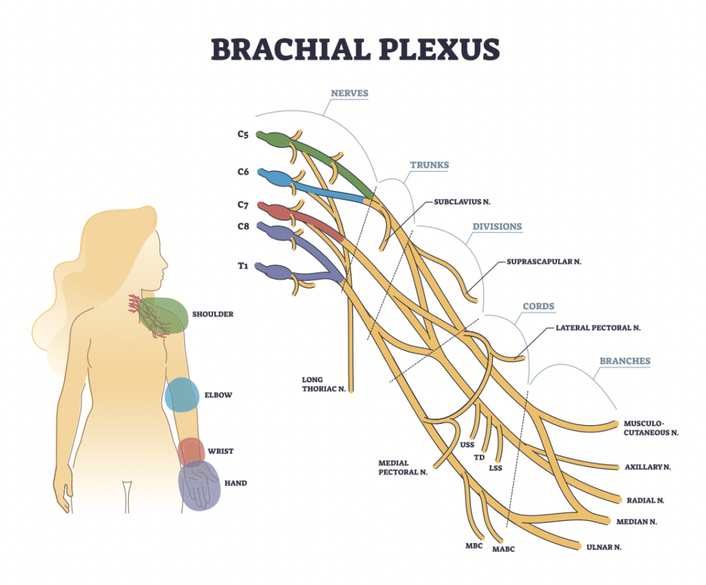
What is the brachial plexus?
The brachial plexus is a group of nerves originating from the cervical and thoracic nerve roots (from C5 to T1). The brachial plexus forms 5 peripheral nerves of the upper extremity, consisting of the musculocutaneous, median, radial, ulnar, and axillary nerves. This group of nerves supplies motor and sensory innervation to the entirety of the upper extremity.
How do injuries happen?
Brachial plexus injuries have multiple mechanisms of injury in infants. These injuries can be caused by compression, traction, stretching, rupture, or avulsion of the nerves of infants during childbirth. Larger infants are at a higher risk of brachial plexus injuries due to the risk of shoulder dystocia (the shoulder of the fetus gets stuck on the pelvis during childbirth). Injuries can also be caused by breech presentation at birth, uterine abnormalities such as uterine fibroids, or the fetus being in a transverse position for a prolonged period of time.
Erb’s palsy vs. Klumpke’s palsy
Erb’s palsy is an upper brachial plexus injury from C5-C6 (sometimes involving C7), while Klumpke’s palsy is a lower brachial plexus injury from C8-T1 (sometimes C7 is involved as well). Erb-Klumpke’s (total paralysis) can also occur if the entirety of the brachial plexus is involved with the injury (C5-T1).
Erb’s palsy causes weakness or paralysis of muscles of the upper arm and shoulder, presenting as the internal rotation of the forearm and flexion of the wrist and fingers with the arm hanging, also called waiter’s tip deformity. Klumpke’s palsy involves weakness or paralysis of the muscles of the forearm and hand, commonly presenting as a “claw hand” with the forearm in supination and flexion of the wrist and fingers.
How are they treated?
Many cases of brachial plexus injuries recover independently with time; however, nonsurgical and surgical treatments are available. Therapy is a common form of treatment that promotes passive range of motion of the shoulder, elbow, wrist, and hand to avoid stiffness of joints. Parents are educated on exercises they can practice with their children at home. Surgery is also an option to repair any rupture present or perform a nerve transfer from another muscle to restore the function of the affected muscles if no progress has been made through the conservative route.
References:
Benjamin, K. (2005). PART 1. Injuries to the brachial plexus. Advances in Neonatal Care, 5 (4), 181-189. doi: 10.1016/j.adnc.2005.03.004.
Brachial plexus injury. Johns Hopkins Medicine. (2022, December 22). https://www.hopkinsmedicine.org/health/conditions-and-diseases/brachial-plexus-injuries
Erb’s palsy (brachial plexus birth palsy) – orthoinfo – AAOS. OrthoInfo. (n.d.). https://orthoinfo.aaos.org/en/diseases–conditions/erbs-palsy-brachial-plexus-birth-palsy/
Klumpke paralysis. Physiopedia. (n.d.). https://www.physio-pedia.com/Klumpke_Paralysis professional, C. C. medical. (n.d.). Shoulder dystocia: Signs, causes, prevention & complications. Cleveland Clinic. https://my.clevelandclinic.org/health/diseases/22311-shoulder-dystocia
More To Read

The Use of Neuromuscular Electrical Stimulation with Upper Extremity Paralysis
The Use of Neuromuscular Electrical Stimulation with Upper Extremity Paralysis By: Mikayla Murphy Martin, R., Johnston, K., & Sadowsky, C. (2012). Neuromuscular electrical stimulation–assisted grasp training and restoration of function in the tetraplegic hand: A case series. The American Journal of Occupational Therapy, 66(4), 471-477. https://doi.org/10.5014/ajot.2012.003004 The Skinny The purpose of the study was to…

Intrinsic Hand Strengthening with Puttycise Tools
We are always looking for ways of the intrinsic hand strengthening. It is easy to overlook the importance of these small but mighty muscles. They are essential to performing functional grasps patterns. They can become weak in a short period of time due to their small size. So, How does intrinsic strengthening work?! The Basics…

Dog Bites to the Hand: What Every Hand Therapist Should Know
What to Expect with a Dog Bite to the Hand for Hand Therapists By: Kathryn Harada Prevalence and Severity:One reason people seek hand therapy is for rehabilitation after an animal bites. In the US alone, 1% of emergency department visits are due to animal bites each year, resulting in 2 to 5 million animal bites…

5+ Common Mallet Finger Splints
Finger orthoses can be tough, and the mallet orthosis is no exception in hand therapy. The protocol for 15 degrees of DIP extension with mallet fingers is tricky to manage while making a common mallet finger splint. Small splints on little fingers are also tricky to get sized just right and with strapping in the…

Sign-up to Get Updates Straight to Your Inbox!
Sign up with us and we will send you regular blog posts on everything hand therapy, notices every time we upload new videos and tutorials, along with handout, protocols, and other useful information.